INTENDED USE
Immunoassay for the in vitro quantitative determination of ferritin in human serum.
Summary
Ferritin is a macromolecule with a molecular weight of at least 440 kD (depending on the iron content) and consists of a protein shell (apoferritin) of 24 subunits and an iron core containing an average of approx. 2500 Fe3+ ions (in liver and spleen ferritin). (1) Ferritin tends to form oligomers, and when it is present in excess in the cells of the storage organs there is a tendency for condensation to semicrystalline hemosiderin to occur in the lysosomes. At least 20 isoferritins can be distinguished with the aid of isoelectric focusing. (2) This microheterogeneity is due to differences in the contents of the acidic H and weakly basic L subunits. The basic isoferritins are responsible for the long-term iron storage function, and are found mainly in the liver, spleen, and bone marrow. (1, 3) Acidic isoferritins are found mainly in the myocardium, placenta, and tumor tissue. They have a lower iron content and presumably function as intermediaries for the transfer of iron in various syntheses (4, 5, 6) The determination of ferritin is a suitable method for ascertaining the iron metabolism situation. Determination of ferritin at the beginning of therapy provides a representative measure of the body’s iron reserves. A storage deficiency in the reticulo-endothelial system (RES) can be detected at a very early stage (7) Clinically, a threshold value of 20 μg/L (ng/mL) has proved useful in the detection of prelatent iron deficiency. This value provides a reliable indication of exhaustion of the iron reserves that can be mobilized for hemoglobin synthesis. Latent iron deficiency is defined as a fall below the 12 μg/L (ng/mL) ferritin threshold. These two values necessitate no further laboratory elucidation, even when the blood picture is still morphologically normal. If the depressed ferritin level is accompanied by hypochromic, microcytal anemia, then manifest iron deficiency is present. (1) When the ferritin level is elevated and the possibility of a distribution disorder can be ruled out, this is a manifestation of iron overloading in the body. 400 μg/L (ng/mL) ferritin is used as the threshold value. Elevated ferritin values are also encountered with the following tumors: acute leukemia, Hodgkin’s disease and carcinoma of the lung, colon, liver and prostate. The determination of ferritin has proved to be of value in liver metastasis. Studies indicate that 76 % of all patients with liver metastasis have ferritin values above 400 μg/L (ng/mL). Reasons for the elevated values could be cell necrosis, blocked erythropoiesis or increased synthesis in tumor tissue. Two monoclonal mouse antibodies - M-4.184 and M-3.170 - are used to form the sandwich complex in the
Test principle
Sandwich principle.
Total duration of assay: 80 minutes.
• Sample, Anti-Ferritin coated microwells and enzyme labeled Anti-Ferritin are combined.
• During the incubation, ferritin presents in the sample is allowed to react simultaneously with the two antibodies, resulting in the ferritin molecules being sandwiched between the solid phase and enzyme-linked antibodies.
• After washing, a complex is generated between the solid phase, the TSH within the sample and enzyme-linked antibodies by immunological reactions.
• Substrate solution is then added and catalyzed by this complex, resulting in a chromogenic reaction. The resulting chromogenic reaction is measured as absorbance.
• The absorbance is proportional to the amount of ferritin in the sample.
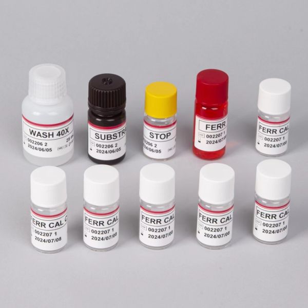
Product details | Description |
Delivery | Within 48 hours |
Packaging Specifications | 8 x 12 strips, 96 wells |
Country Of Origin | China |
Manufacturer | 18 months |
Preservation method | 2℃-8℃ |
Specimen | Whole blood |
Assification | class1 |
Type | Elisa Test Kit |