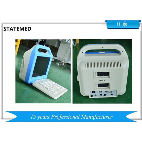
Veterinary Handheld Ultrasound Scanner For Cow Pregnancy Test
STT3-VUS29
1. Technical Parameters
Scanning mode: Electron convex array scanning,
Electron linear array scanning
Electron convex array
Five display format: B, B+B, 4B, B+M, M
Image magnification: ×1.0, ×1.2, ×1.3, ×1.5, ×1.6, ×1.8, ×2.0
L-zoom: real-time partial amplification
Depth of detection: ≥180mm
Image grey scale: 256 levels; 4-stageγcorrection
Frame: 3-stage frame function
Focus: Dynamic digital electronic focus point by point
Gain control: Full gain and 8-segment TGC
Probe Conversion: probe automatic identification
Frequency for Convex : 2.0MHz, 2.5 MHz, 3.0 MHz, 3.5 MHz, 4.0 MHz, 4.5 MHz, 5.0 MHz
Note: Hospital name, age, sex; organ automatic annotation function; Partial and full-screen character annotation function.
Display: English/Chinese conversion function; Fixed characters, real-time colock, calendar display.
Veterinary Body Mark : 5 kinds
Puncture function: puncture guide line displayed in B mode.
Measurement function: Bovine: BPD , CRL; Equine :BPD, GSD; Ovine: BPD, CRL; Canine: CRL, BD;
Feline: BD, HD. Image display: image freeze, real-time, up/down, left/right, black/white reverse.
Cine loop: 512-frame cine loop and storage.
Image storage: store 16 thousand image by USB flash.
Printing function: connect with laser printer or video printer
Power saving function
Output interface: USB interface, standard mini port (and trapezoidal interface), video printer interface
Print: machine can connect any video printers directly.
Equipment for the injection shell structure. The host uses non-frequency transformer switching power supply, and adopt to the programmable devices and surface mount technology (SMT). The whole machine with high degree of integration, excellent performance.
2. Main Technical Features
2. 2. 1 performance of standard probe
2. 2. 1. 1 R=60 the center frequency of convex array probe: 3.5Mhz
2. 2. 1. 2 investigation depth: >=180mm
2. 2. 1. 3 Vertical resolution:<=1mm(depth<=130mm)
<=2mm(130mm<depth<=170mm)
2. 2. 1. 4 Lateral resolution:<=2mm(depth<=130mm)
<=3mm(130mm<depth<=160mm)
2. 2. 1. 5 Geometric position accuracy: Portrait<=5%; cross wise<=10%;
Dead zone: <=4mm
Slice thickness : <=5mm
Pupil deviation:<=+-20%
M mode display error: <=5%
Main configure probe 3.5Mhz(R=60) the frequency of convex array probe: 2.0Mhz, 2.5Mhz, 3.5Mhz, 3.5Mhz, 4.0Mhz, 4.5Mhz, 5.0Mhz. The working frequency performance requirements listed in Table 1;
Table 1: B ultrasonic diagnostic performance requirements file
performance | Probe style and standard frequency | |||
Standard frequency of probe | 3.5 MHz | 6.5MHz | ||
Random error of frequency | Deviation of Sound frequency and standard frequency should ≤±15% | |||
Probe | linear, R≥60mmconvex | linear(linear), R≥60mm convex | R<60mm convex
| |
Investigation depth,mm | ≥180 | ≥70 | ≥60 | |
Axial resolution,mm | ≤2 (depth≤80) ≤3 (80<depth≤130) | ≤1 (depth≤50) | ≤1 (depth≤40) | |
Lateral resolution,mm | ≤3 (depth≤80) ≤4 (80<depth≤130) | ≤2 (depth≤40) | ≤2 (depth≤30) | |
Geometric position accuracy,% | crosswise≤10 Length wises≤5 | crosswise≤5 Length wises≤5 | crosswise≤10 Length wises≤5 | |
Dead zone,mm | ≤4 | ≤3 | ≤4 | |
Slice thickness,mm | ≤9 | ≤5 | ≤5 | |
Pupil error,% | ≤±20 | ≤±20 | ≤±20 | |
M mode time error,% | ≤5 | -- | -- | |
M mode distance error,% | ≤5 | -- | -- | |
Related Ultrasound machines