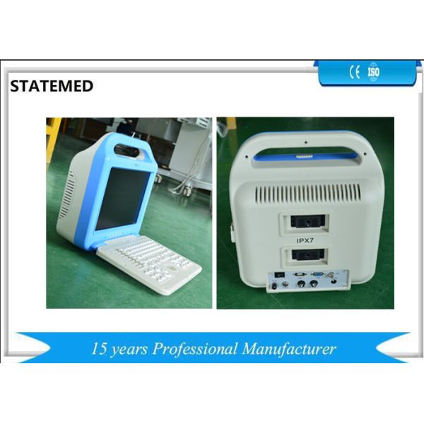
Portable Ultrasound Ce Approved Veterinary Use Full digital Ultrasound Scanner
1. Operation functions of Equipment
● Four display format: B,B+B,4B,B+M,M; Image grey scale: 256 levels.
● Freezing, real time, selective enlargement, upwards/downwards and left/right turnover of the image can be realized.
● 8 stage TGC adjustment.
● 5 types of body mark.(Swine, Dog,Equine, Bovine, Sheep)
● Full-screen character annotation.
● Fixed character, real time clock and calendar.
● Electron focusing, free combination of focus.
● Various probe for choice, enough to meet requirement of different animals
● Frequency Conversion: 2.0MHz – 8.0 MHz.
● Image magnification: ×1.0,×1.2,×1.5,×2.0. the utmost depth of detection is 240mm.
● Measuring: distance, perimeter, area, volume, pregnant weeks etc. Heart rate and slope can be measured under M mode.
● Built-in DSTN Processor, PAL-D and VGA video transmission, external screen monitor, colorful screen, video tape recorder, video image printer etc.
● Movie replay function, uninterrupted replay or step forward/backward replay for 256 images can be realized.
● Having the function of “flash in”,“flash out”, “dream in”, “dream out” and “USB”.
● With the optional function of 4 segments frame correlation.
2. Technical Specification
The main functions of the main probe
2. 1. 1 The frequency of Electronic Convex Array probe is 3.5MHz(R=60)
2. 1. 2 Depth display: ≥190mm
2. 1. 3Resolution (vertical direction): ≤1mm (depth ≤130mm)
≤2mm(130mm<depth ≤160mm)
2. 1. 4 Resolution (cross direction) ≤2mm (depth ≤130mm)
≤3mm(130mm<depth ≤160mm)
2. 1. 5 Precision of geometric position :cross direction≤10%; vertical direction≤ 5%
2. 1. 6 Blind Zone(mm): ≤4mm
2. 1. 7 The conversional frequency points of the main probe (3.5MHz Convex Array probe) are: 2.0MHz﹑3.5 MHz﹑4.5 MHz﹑5.0 MHz﹑5.5 MHz﹑6.5 MHz﹑7.5 MHz , the detailed requirements of the conversion frequency points are as follows:
The frequency of probe (MHz) | f≤2.5 | 2.5<f≤4.0 | 4.0<f≤5.0 | 5.0<f≤10 | |
Type of probe | liner array , R≥60mm convex array | liner array , R≥60mm convex array
| liner array , R≥60mm convex array
| Superficial R≥60mm convex array
| (intra-cavityprobe ) R<60mm convex Array) |
Resolution (Lateral direction) mm | ≤3 (depth)≤130) ≤4(130<depth≤160) | ≤2 (depth≤130) ≤3 (130<depth≤160) | ≤2 (depth≤60) | ≤1 (depth≤60) | ≤1 (depth≤40) |
Resolution (Vertical direction) mm | ≤1 (depth≤130) ≤2 (130<depth≤170) | ≤1 (depth≤130) ≤2 (130<depth≤170) | ≤1 (depth≤80) | ≤1 (depth≤80) | ≤1 (depth≤40) |
Blind zone,mm | ≤4 | ≤4 | ≤3 | ≤3 | ≤5 |
Utmost depth display,mm | ≥190 | ≥190 | ≥120 | ≥80 | ≥60 |
Precision of geometric position,% | Lateral≤10 Vertical direction≤5 | Lateral≤10 Vertical direction≤5 | Lateral≤10 Vertical direction≤5 | Lateral≤5 Vertical direction ≤5 | Lateral≤10 Vertical direction≤5 |
3. Power
When AC 220V±22V or 110V±11V is changed , the equipment can also be operated as usual.
4. Working time
The continental working time for the Ultrasonic Diagnosis Equipment is more than 8 hours.
5. Selective configurations
The medical image work station can be connected with the ultrasonic if the function index of the former satisfies the requirements of the standard.
6. Safety
There are 60 items of Security Detection Project adapts to the portable Ultrasound in the appendix A.
If any questions,please feel free to contact me!