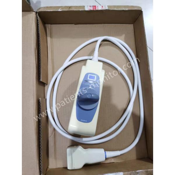
L12-5 50mm PHiliphhilip Linear Array Transducer For EPIQ 7, EPIQ 5, Affiniti 70, Affiniti 50, IU22, HD7 XE, HD9, HD11 XE HD15, and CX50 Ultrasound Systems
The PHiliphhilip L12-5 50mm Linear Array Explora Transducer has a 5 to 12 MHz operating frequency range and is suitable for Abdominal, Pediatric Hips, Small Parts, and Vascular applications. The PHiliphhilip L12-5 50mm Linear Array Explora Transducer are compatible with PHiliphhilip EPIQ 7, EPIQ 5, Affiniti 70, Affiniti 50, iU22, HD7 XE, HD9, HD11 XE, HD15, and CX50 Ultrasound Systems
Technology | / |
Frequency range | 12-5 MHz |
Aperture | 50mm- |
Volume of field of view | / |
Applications | Breast, thyroid and superficial small parts; musculoskeletal tendon, abdomen bowel, and vascular applications |
Image Fusion Navigation capable | Yes |
Number of elements | 256 |
Array Type | Linear |
Modes | 2D, steerable pulsed wave and color Doppler, color power angio (CPA), SonoCT, variable XRES, and multivariate harmonic imaging |
Biopsy capable | Yes |
PHiliphhilipysical dimensions | Cable length: 78 in/198 cm |
• Fine pitch, 256 element, high resolution linear array
• 12 to 5 MHz extended operating frequency range
• 10° of trapezoidal imaging
• Steerable pulsed wave, color Doppler, and Color Power Angio, SonoCT, XRES, and harmonic imaging
• Intelligent Doppler flow optimization
• Cable length: 78 in/198 cm
• Breast, thyroid and superficial small parts; musculoskeletal tendon, abdomen bowel, and vascular applications
• Biopsy guide available
• AutoSCAN imaging is available on L12-5
About Transducer
The transducer you choose is the most important factor affecting image quality. The best imaging cannot be obtained without the correct transducer. The system is optimized for use according to the transducer you choose.
The system limits the patient's exposure temperature to 43 degrees Celsius and limits the acoustic output value to the respective U.S. Food and Drug Administration limits. The power supply protection circuit prevents overcurrent conditions. If the power monitor protection circuit detects an overcurrent situation, it will immediately cut off the sensor's drive voltage to prevent the sensor surface from overheating and limit the sound output. The verification of the power protection circuit is completed under normal system operation.
To limit the potential hazards when scanning neonatal, pediatric, and medication patients, try to minimize the time spent imaging at temperatures above 41°C (106°F).
When the system is not in use, press Freeze to stop ultrasound transmission
It is important to understand how to connect, disconnect, and select sensors, and how to use the cable management system on the system cart.
The system includes a socket for imaging transducers and a socket for pulsed or continuous wave Doppler probes. Two sockets can be occupied at the same time, but only one transducer can be activated at a time.
On the optional cart, when the transducer is not in use, store it in one of the transducer holders on the system cart. When the transducer is not connected, place the connector cover on the connector and store the connector in a bracket on the back of the cart. Always use the cable management system on the optional cart to prevent the cables from being stepped on or crushed by the cart wheels.