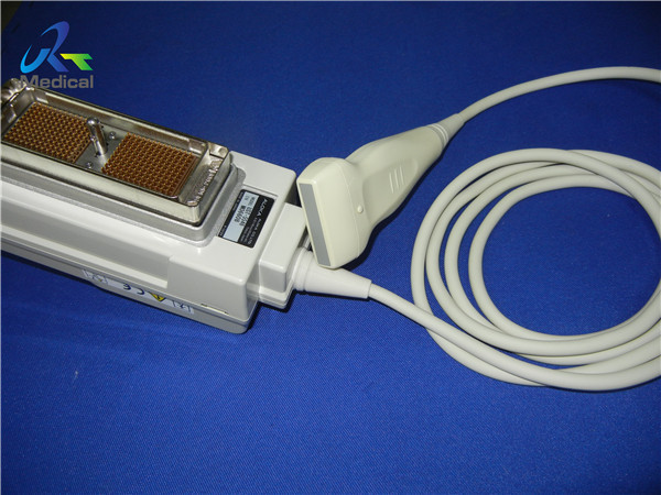
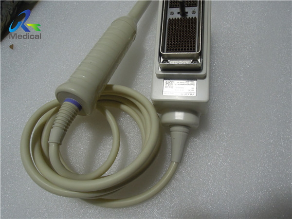
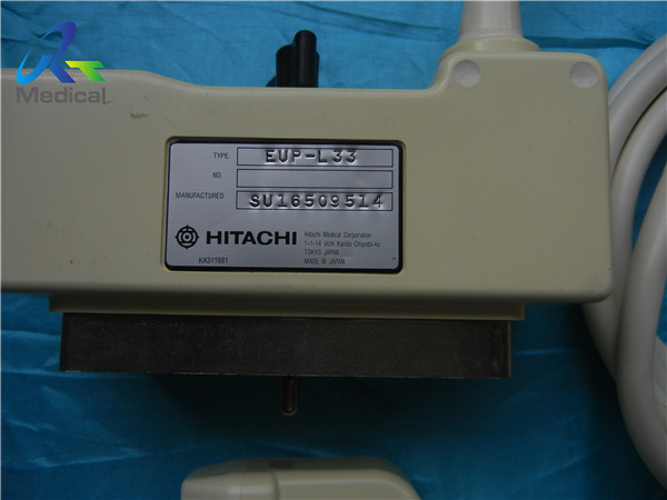
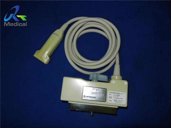
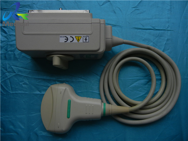
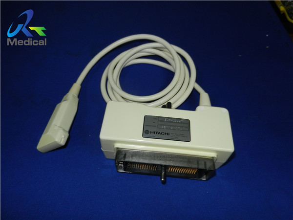
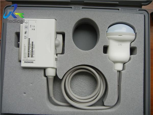
Toshiba PVT-661VT 10mm Ultrasound Machine Transducer Probes Endovaginal Diagnostic Equipment
PVT-661VT 10mm Ultrasound Scanner Probe For Endovaginal Diagnostic Equipment With Enhanced Imaging Capabilities
1. Type: Endovaginal
2. Frequency: 3-9MHz
3. Compatible system: Aplio 50 SSA-700A, Aplio SSA-750A, Xario SSA-660A
4. Condition: original, in good working condition
5. With 60 days warranty
Other Toshiba probes we can offer:
Brand | Model | Compatible System |
Toshiba/Canon | PLF-805ST | SSA-340A&SSA-350A |
Toshiba/Canon | PLM-1204AT | PowerVision 6000 SSA-370A/ Nemio 17 SSA-550A/ Xario SSA-660A |
Toshiba/Canon | PLM-703AT | Powervision 6000 and Nemio |
Toshiba/Canon | PLM-805AT | PowerVision 6000 SSA-370A/ Nemio 17 SSA-550A |
Toshiba/Canon | PLT-1005BT | Aplio 300/ Aplio 400/ Aplio 500 |
Toshiba/Canon | PLT-1204AT | Aplio 50 SSA-700A/ Aplio SSA-750A/ Xario Series |
Toshiba/Canon | PLT-604AT | Xario Series / Aplio 50 SSA-700A/ Aplio SSA-750A |
Toshiba/Canon | PLT-704AT | Aplio 50 SSA-700A/ SSA-750A/ Xario |
Toshiba/Canon | PLT-704SBT | Xario SSA-660A |
Toshiba/Canon | PLT-805AT | SSA-700A/ Aplio SSA-750A/ Aplio SSA-770A/ Xario SSA-660A |
Toshiba/Canon | PLU-1204BT | Xario 100/Xario 200 |
Toshiba/Canon | PSM-25AT | PowerVision 6000 SSA-370A/ Nemio 17 SSA-550A |
Toshiba/Canon | PSM-37CT | PowerVision 6000 SSA-370A/ Nemio 17 SSA-550A |
Toshiba/Canon | PST-25ST | Viamo |
Toshiba/Canon | PST-30BT | Xario & Aplio Series |
Toshiba/Canon | PST-30BT | Aplio 50 SSA-700A/ Aplio SSA-750A/ Artida SSH-880CV/ Xario SSA-660A |
Toshiba/Canon | PSU-25BT(5S1) | Xario 100/ Xario 200 |
Toshiba/Canon | PSU-30BT(5S2) | Xario 100/ Xario 200 |
Toshiba/Canon | PVE-382M | 77B/90A/100A/240A/250A/TOSBEE |
Toshiba/Canon | PVF-375MT | SSH-140A/ SSA-270A/ SSA-340A |
Toshiba/Canon | PVM-375AT | PV 6000 SSA-370A/ Nemio 17 SSA-550A/ Nemio XG SSA-580A/ Nemio 30/ SSA-370A |
Toshiba/Canon | PVM-621VT | PowerVision 6000 SSA-370A/ Nemio 17 SSA-550A |
Toshiba/Canon | PVM-651VT | PowerVision 6000 SSA-370A/ Nemio 17 SSA-550A |
Toshiba/Canon | PVT-375BT | Aplio 50 SSA-700A/ Aplio SSA-750A/ Aplio SSA-770A/ Aplio XG SSA-790A/ Xario SSA-660A/ SSA-680A |
Toshiba/Canon | PVT-375BT | SSA-700A/SSA-750A/ Aplio SSA-770A/SSA-790A/SSA-660A/ SSA-680A |
Toshiba/Canon | PVT-382BT | Aplio XG/Aplio MX/Xario XG/Xario/Viamo (2.0 and above)/Aplio 500/Aplio 300 |
Toshiba/Canon | PVT-575MV | Xario SSA-660A/ Aplio XG |
Toshiba/Canon | PVT-661VT | Aplio 50 SSA-700A/ Aplio SSA-750A/ Xario SSA-660A |
Toshiba/Canon | PVT-674BT | Aplio 50 SSA-700A/ Aplio SSA-750A |
Toshiba/Canon | PVT-675MV | Xario 100/ Xario 200/ Aplio MX |
Toshiba/Canon | PVT-681MV | Aplio 300/ Aplio 400/ Aplio 500/ Xario XG |
Toshiba/Canon | PVT-712BT | Aplio series/ Xario series |
Toshiba/Canon | PVU-781VT | Xario |
Knowledge point
Color-flow imaging is an advanced iteration of Doppler ultrasound technology, providing a sophisticated means to visualize blood flow within the body. In a process akin to duplex ultrasound, this technique employs color to accentuate the direction of blood flow, offering a dynamic representation of vascular activity. Vessels conducting blood exhibit a red hue for flow in one direction and a blue hue for flow in the opposite direction. A color scale is employed to convey the speed of the flow, enabling a comprehensive assessment of circulatory dynamics.
This imaging modality excels in elucidating the anatomical structure and behavior of large blood vessels. By visually presenting the direction, speed, and range of blood flow, color-flow imaging facilitates the identification of potential blood flow disorders and abnormal pathways. The color-coded depiction allows for a clear and intuitive interpretation of circulatory patterns.
The velocity of blood flow is a critical parameter influencing the reflection frequency, and this is effectively captured by the amplitude beam in spectral Doppler. High blood flow velocity corresponds to a heightened amplitude on the spectrum curve, while slower blood flow is reflected by a lower amplitude. This amplitude information serves as a precise metric for calculating the velocity of blood flow, contributing to the accuracy of diagnostic assessments.
Color Doppler images are not only informative but also visually engaging, presenting different levels of brilliance to convey varying flow velocities. This nuanced display enhances the interpretability of the images, providing healthcare professionals with a comprehensive and intuitive tool for assessing blood flow dynamics. In summary, color-flow imaging stands as a valuable enhancement to Doppler ultrasound, offering detailed insights into circulatory patterns and aiding in the identification of potential abnormalities.