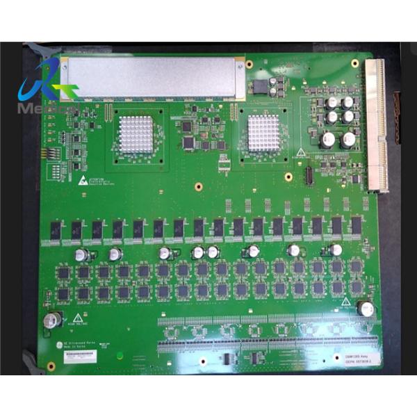
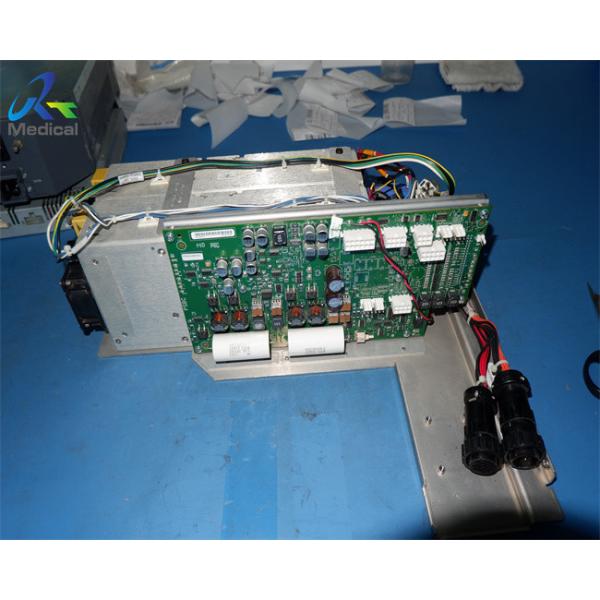
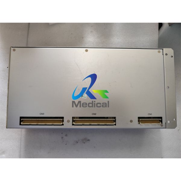
GE Logiq P7 P9 COM module R2/R2.5 Ultrasound Machine Part Medical Use 6324558-4
1. Part number: 6324558-4
2. Compatible system: GE Logiq P7 P9 R2/R2.5
3. New original
4. Warranty: 90 days
5. Lead time: 2-4 days
Outright/exchange/repair is available for ultrasound parts/Assy.
Other ultrasound parts that we can offer:
No. | Model | Part Number | Description | Version |
1 | Logiq P7 P9 | 5501560-141 | LCD | R1-R2 |
2 | Logiq P7 P9 | 7501560-2 | LCD | R3 |
3 | Logiq P7 P9 | 5503745 | MARU MBP PWA main board | Generic version |
4 | Logiq P7 P9 | 5504162-6 | Probe interface board | Generic version |
5 | Logiq P7 P9 | 5372959 | DC4D ASSY 4D Board | Generic version |
6 | Logiq P7 P9 | 5494666-4 | MCB PWA | R1 |
| Logiq P7 P9 | 6494666-2 | MCB-H Assy | R2/R2.5 |
| Logiq P7 P9 | 7494666 | MCB-R3 Assy | R3 |
7 | Logiq P7 P9 | 5554921-4 | IOBOX Assy | R1 |
| Logiq P7 P9 | 5991830-3 | IOBOX-B Assy | R2/R2.5/R3 |
8 | Logiq P7 P9 | 5552220-2 | MDC Assy | R1/R2/R2.5 |
| Logiq P7 P9 | 5552220-3 | MDC-R3 Assy | R3 |
9 | Logiq P7 P9 | 6324558-4 | COM module | R2/R2.5 |
| Logiq P7 P9 | 7324558 |
| R2/R2.5 |
| Logiq P7 P9 | 7324560-2 |
| R3 |
10 | Logiq P7 P9 | 5551110-2 | MIO-2 PWA | R1 |
| Logiq P7 P9 | 6426420-2 | MIO-B PWA | R2/R2.5 |
11 | Logiq P7 P9 | 5737480-4 | Artesyn LP63 ACDC Assy for FRU | R2/R2.5/R3 |
12 | Logiq P7 P9 | 5554918 | Track Ball | R1 |
13 | Logiq P7 P9 | 9554918 | Track Ball | R2-R4.5 |
Knowledge Point
What is Sonography?
Sonography, also called ultrasound imaging or ultrasound scanning, uses high frequency sound waves to obtain a real time image of the inside of the body. The image shows the movement of blood and organs and the structure of internal tissues. Traditionally, a sonogram will show a flat presentation of the examined region, but three dimensional (3D) images are also possible, as are 4D pictures, which show 3D images in motion. The picture is called an ultrasound or a sonogram.
Doppler sonography, or Doppler ultrasound, is a medical imaging technique that uses ultrasound enhanced by the Doppler effect. The Doppler effect generally uses a change in the pitch of sound waves that are measured by an ultrasound-receiving device such as a transducer. Colored Doppler images often provide helpful information about the flow and movement of blood and inner areas of the body. This technique is commonly used during pregnancy to provide detailed images of a developing unborn baby. Other uses for Doppler sonography may include visual examinations of tendons, joints and muscles as well as internal organs.