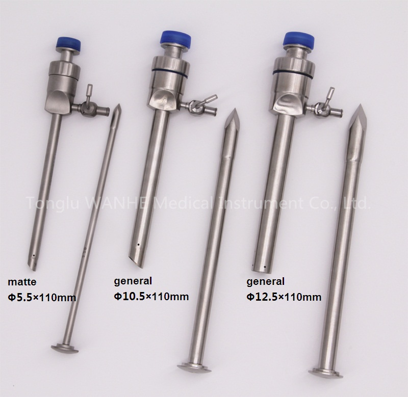
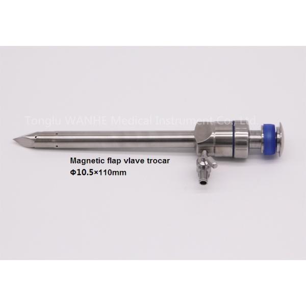
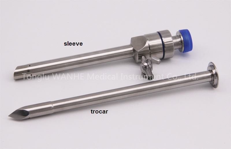
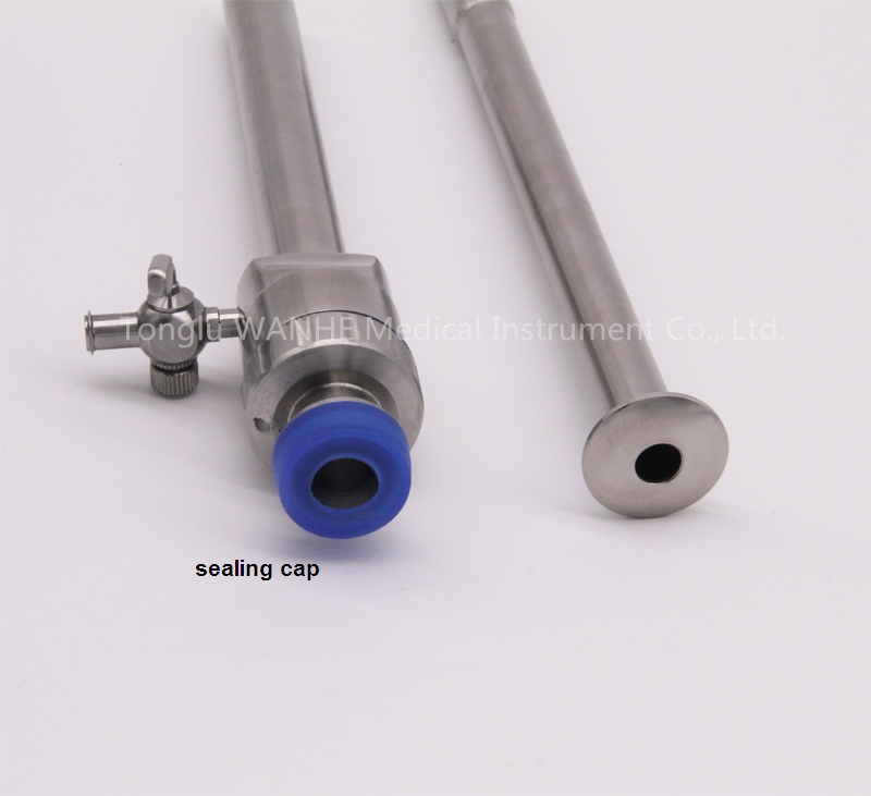
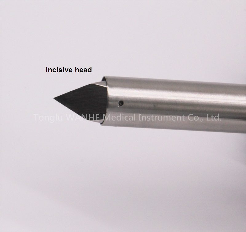
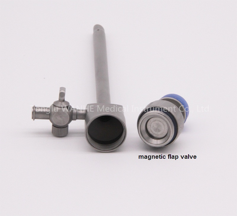
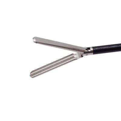
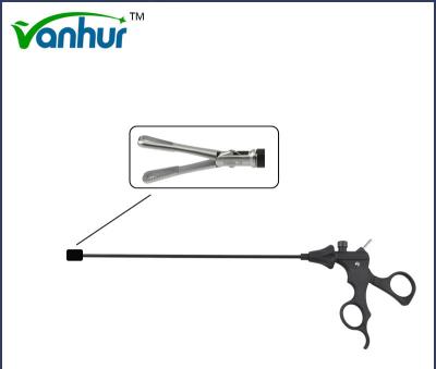
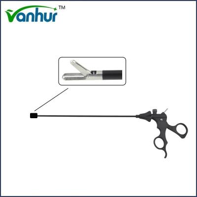
FDA Certified Steel Laparoscopic Instrument with Reusable Magnetic Flap Valve Trocar
1. Trocar Details:
Model | Name | Specifications |
HF2016.12 | Magnetic flap valve trocar | Φ5.5mm |
HF2016.13 | Magnetic flap valve trocar | Φ10.5mm |
HF2016.14 | Magnetic flap valve trocar | Φ11mm |
HF2016.15 | Magnetic flap valve trocar | Φ6mm |
HF2016.16 | Magnetic flap valve trocar | Φ12.5mm |
HF2016.20 | Magnetic flap valve trocar with protection, bloodless | Φ5.5mm |
HF2016.21 | Magnetic flap valve trocar with protection, bloodless | Φ6mm |
HF2016.22 | Magnetic flap valve trocar with protection, bloodless | Φ10.5mm |
HF2016.23 | Magnetic flap valve trocar with protection, bloodless | Φ11mm |
HF2016.38 | Magnetic flap valve trocar with protection, bloodless | Φ12.5mm |
HF2016.17 | Magnetic flap valve trocar with retaining thread | Φ5.5mm |
HF2016.18 | Magnetic flap valve trocar with retaining thread | Φ10.5mm |
HF2016.24 | Magnetic flap valve trocar with retaining thread | Φ6mm |
HF2016.25 | Magnetic flap valve trocar with retaining thread | Φ12.5mm |
HF2016.35 | Magnetic flap valve trocar with retaining thread | Φ11.5mm |
HF2016.30 | Magnetic flap valve trocar with protection, retaining thread | Φ5.5mm |
HF2016.31 | Magnetic flap valve trocar with protection, retaining thread | Φ10.5mm |
HF2016.32 | Magnetic flap valve trocar with protection, retaining thread | Φ6mm |
HF2016.33 | Magnetic flap valve trocar with protection, retaining thread | Φ12.5mm |
HF2016.34 | Magnetic flap valve trocar with protection, retaining thread | Φ11mm |
HF2016.65 | Trumpet trocar | Φ10.5mm |
HF2016.66 | Trumpet trocar | Φ5.5mm |
HF2016.67 | Trumpet trocar with protection | Φ10.5mm |
HF2016.68 | Trumpet trocar with protection | Φ5.5mm |
HF2016.5 | Cross-type trocar | Φ10.5mm |
HF2016.6 | Cross-type trocar | Φ5.5mm |
HF2016.8 | Cross-type trocar | Φ11mm |
HF2016.9 | Cross-type trocar | Φ12.5mm |
HF2016.10 | Cross-type trocar | Φ6mm |
HF2016.3 | Cross-type trocar with protection | Φ10.5mm |
HF2016.4 | Cross-type trocar with protection | Φ5.5mm |
HF2016.7 | Cross-type trocar with protection | Φ11mm |
HF2016.28 | Cross-type trocar with protection | Φ6mm |
HF2016.29 | Cross-type trocar with protection | Φ12.5mm |
HF2016.3A | Cross-type trocar with retaining thread | Φ10.5mm |
HF2016.4A | Cross-type trocar with retaining thread | Φ5.5mm |
HF2016.36 | Cross-type trocar with protection, retaining thread | Φ10.5mm |
HF2016.37 | Cross-type trocar with protection, retaining thread | Φ5.5mm |
HF2017.1 | Adaptor/Reducer | Φ10.5-5.5mm |
HF2017.2 | Adaptor/Reducer, stainless, long tube | Φ10.5-5.5mm |
HF2017.4 | Adaptor/Reducer, stainless, long tube | Φ5-3mm |
HF2017.5 | Adaptor/Reducer, plastic | Φ10.5-5.5mm |
HF6002.1 | Sealing cap | Φ5mm |
HF6002.2 | Sealing cap | Φ10.5mm |
HF6002.3 | Sealing cap | Φ12.5mm |
HF6001.1 | Cross-type membrance valve | Φ5mm |
HF6001.2 | Cross-type membrance valve | Φ10mm |
HF6003.2 | O-ring | Φ10.5mm |
HF6003.3 | O-ring | Φ5.5mm |
Package detail: | Poly bag and special shockproof paper box. |
Delivery detail: | By air |
FAQ
Laparoscopic surgery has many advantages over traditional surgery, which are mainly reflected in the following aspects:
Less trauma: Laparoscopic surgery is performed by making several small holes in the abdominal wall, and the incision is significantly smaller than that of traditional laparotomy. The incision of traditional surgery can reach more than ten to twenty centimeters, while the incision of laparoscopic surgery is only about 0.5 to 1 centimeter.
Quick recovery: Due to the smaller trauma, the patient has less pain after surgery, the recovery time is short, and the hospitalization time is also shortened accordingly. The patient can eat semi-liquid food and get out of bed the next day after surgery, and can resume normal life and work after one week.
Fewer complications: Laparoscopic surgery has less trauma and less interference with the abdominal cavity, and the chance of intestinal adhesion after surgery is lower. In addition, the incidence of postoperative complications is low, such as reduced intestinal adhesion infection.
Aesthetics: The incision of laparoscopic surgery is hidden, leaving no obvious scars, local beauty, small postoperative scars, and extremely beautiful.
Less bleeding: Laparoscopic surgery has less bleeding, short operation time, and less intraoperative bleeding.
However, laparoscopic surgery also has its limitations and disadvantages:
High technical requirements: Laparoscopic surgery has high technical requirements for doctors, and the operation is difficult, requiring high suturing skills and precise operation capabilities.
Expensive equipment: Laparoscopic equipment is expensive, which increases the cost of surgery.
Possible omission of lesions: Since the tumor site is felt by instruments, it is not as sensitive and intuitive as direct contact with hands, and the tumor positioning is not as good as open surgery. If the location of polyps and tumors cannot be determined by laparoscopic surgical instruments, it may be necessary to accurately locate them by other means before completing the operation.
In summary, laparoscopic surgery has significant advantages in terms of less trauma, faster recovery, and fewer complications, but it also has disadvantages such as high technical requirements, expensive equipment, and possible omission of lesions. When choosing a surgical method, it is necessary to consider the specific situation and the patient's condition comprehensively.
The specific differences in postoperative recovery time between laparoscopic surgery and traditional surgery are mainly reflected in the following aspects:
Discharge time:
Laparoscopic surgery usually allows patients to be discharged earlier. For example, after single-port laparoscopic surgery, patients can move within 6 hours after surgery and be discharged within 1-2 days. In contrast, patients often need about a week to be discharged after traditional surgery.
Time to resume basic activities:
After laparoscopic surgery, patients can resume basic activities within 2-3 days after surgery. Traditional surgery requires at least 2 weeks to resume basic activities.
Time to complete recovery:
The time to complete recovery from laparoscopic surgery varies depending on the type of surgery and the patient's physical condition. For minor surgeries, such as laparoscopic cholecystectomy or ovarian cystectomy, patients can return to normal about 20 days after surgery. For more complex surgeries, such as liver cancer resection or radical gastrectomy, the recovery time will be longer, which may take 1-3 months or even longer.
The recovery time for traditional surgery is usually longer, especially for surgeries with large wounds, where patients may need longer time to recover.
Time for wound healing:
Laparoscopic surgery wounds can generally heal completely within 5-7 days. Traditional surgery wound healing time is usually longer.
Laparoscopic surgery has obvious advantages in postoperative recovery time, especially in terms of discharge time, time to resume basic activities, and time to wound healing.
Laparoscopic surgery is a surgical technique that uses a miniature camera for internal vision. This surgical method is performed through a laparoscope and its related instruments, using a cold light source to provide illumination, inserting a laparoscope lens (3-10mm in diameter) into the abdominal cavity, and using digital camera technology to transmit the image captured by the laparoscope lens through optical fibers to the post-stage signal processing system, and display it in real time on a dedicated monitor.
Technology and Equipment
The 4K laparoscopic system provides a higher-definition surgical field of view and more delicate detail resolution, and has significant advantages in grasping the membranous anatomical level, identifying fine blood vessels or nerves, and identifying the boundaries of lymph node dissection. This high-resolution field of view improves the surgeon's sense of the surgical field of view and promotes the further development of laparoscopic surgery technology.
This system sets a benchmark for quality and versatility for multidisciplinary use, can assist in a wide range of surgical applications, and provide precise, reliable and intuitive operation.
Disposable high-frequency surgical instruments can meet the needs of various surgical scenarios, including disposable single-action fine-tooth grasping forceps, double-action fine-tooth grasping forceps, corrugated grasping forceps, blunt-head grasping forceps, gallbladder grasping forceps, mouse-type inverted tooth grasping forceps and duckbill grasping forceps.
The new laparoscopic surgical instrument bag has achieved remarkable results in preventing postoperative infection, improving work efficiency and ensuring the safe use of instruments.
This device adopts the end-decoupled wire-driven multi-degree-of-freedom surgical instrument design, which enhances the flexibility of doctors' operations; the folding distal centering instrument operating arm also improves the accuracy of the operation.
Impact
The application of these technologies and equipment in laparoscopic surgery has significantly improved the surgical effect and safety:
The high-definition field of view provided by the 4K laparoscope system enables surgeons to more clearly identify and operate subtle structures, thereby reducing the risk of accidental injury and improving the success rate of surgery.
The use of disposable high-frequency surgical instruments reduces the risk of cross-infection, meets the needs of various surgical scenarios, and improves the adaptability and flexibility of surgery.
The improved laparoscopic surgical instrument bag performs well in preventing postoperative infection, ensuring the safe use of instruments and reducing the risk of surgical complications.
The results of the comparison of laparoscopic surgery with traditional surgery in terms of cost-effectiveness are inconsistent, depending on the type of surgery and study design.
Laparoscopic radical resection of colorectal cancer is considered to be an effective method with less trauma, less bleeding, and faster recovery compared with traditional open surgery, and is worthy of clinical promotion and application. Although no specific cost-effectiveness data are provided, this description implies that laparoscopic surgery may have higher value in some aspects.
The results of the comparison of laparoscopic surgery with traditional surgery in terms of cost-effectiveness vary depending on the type of surgery and study design. In some cases, such as the treatment of gastroesophageal reflux disease, laparoscopic surgery shows better cost-effectiveness; while in other cases, such as the treatment of early hepatocellular carcinoma, laparoscopic surgery is more expensive and may not have the best cost-effectiveness.
Although laparoscopic surgery has the advantages of less trauma and faster recovery, there are still certain complications and types of lesions that may be missed. The following are some common missed lesions and their prevention and detection methods:
Ureteral injury: In gynecological laparoscopic surgery, ureteral injury is one of the more common complications. To prevent this, the cystic duct, common bile duct, and common hepatic duct can be fully dissected and exposed during the operation before clamping and cutting the cystic duct.
Bladder injury: Bladder injury is also a common problem in laparoscopic surgery. The position relationship of the three ducts should be clarified by angiography during the operation to reduce the risk of accidental injury.
Pelvic infection: Pelvic infection is another common complication of laparoscopic surgery. For prevention, the pneumoperitoneum pressure should be strictly controlled between 10 and 15 mmHg during the operation, and strict laparoscopic operation training should be performed.
Bile duct injury: In laparoscopic cholecystectomy, bile duct injury may cause bile leakage. For prevention, the operator needs to undergo strict laparoscopic operation training. Each case should be fully dissected and exposed to identify the relationship between the cystic duct, common bile duct, and common hepatic duct before clamping and cutting the cystic duct.
Subcutaneous emphysema or gas embolism: These complications can cause local pain, bleeding, etc. After surgery, you can use blood-activating drugs under the guidance of a doctor to relieve these symptoms.
In order to discover these lesions that may be missed, the following follow-up examinations can be performed:
Postoperative imaging examinations: such as ultrasound examinations, CT scans, etc., can help to promptly detect problems such as ureteral injury, bladder injury, and bile duct injury.
Blood tests: such as arterial blood gas analysis and blood oxygen saturation testing, can detect problems such as abnormal cardiopulmonary function at an early stage.
Regular follow-up: Regular follow-up after surgery to promptly detect and treat postoperative complications such as pelvic infection, subcutaneous emphysema or gas embolism.
For more photos and details please contact us:
Tonglu Wanhe Medical Instruments Co., Ltd.