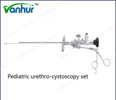
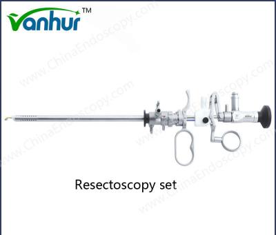
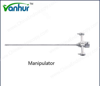
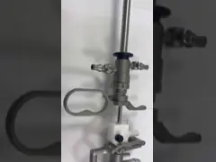
OEM Acceptable Surgical Hysteroscopy Instruments Electrode Spike for Resectoscopy
1 Introduction:
If you are looking for minimally invasive surgery medical instruments with good quality, competitive price and reliable service. Wanhe medical is manufaturing these for you. We provide general and professional laparoscopic instruments with CE, FDA approved.
2 Specifications
Adopt 3Cr13, 304, 630 stainless steel material
Tough construction
Corrosion resistant
High durability
Safety application
3 Packing & Shipping:
| Package detail: |
Poly bag and special shockproof paper box. |
| Delivery detail: |
By air |
FAQ
What is the surgical preparation and preoperative planning for urinary surgical instruments?
The preparation and preoperative planning of urinary surgical procedures are important links to ensure the smooth progress of the operation and improve the patient's postoperative recovery. The following are detailed preparation and planning steps:
Surgical preparation
Environment and equipment preparation
The operation should be performed in a certified sterile operating room with a new, clean bandage.
Ensure that all necessary medical equipment and instruments (such as various types of blades, forceps, scissors and tweezers, optical equipment, etc.) are ready.
Patient assessment and examination
Perform a comprehensive physical examination, including blood routine, urine routine, liver and kidney function, electrocardiogram, chest X-ray, B-ultrasound, etc.
Perform CT or MRI imaging on the patient to evaluate the spatial relationship between the kidney and the stone for preoperative planning.
Understand the medical history and perform a physical examination, especially the evaluation of complications such as urinary tract infection or acute renal failure.
Anesthesia plan formulation
Choose general anesthesia or spinal anesthesia according to the type of surgery, and discuss the specific anesthesia plan with the anesthesiologist.
During the induction of general anesthesia, a urinary catheter is placed to improve the patient's urethral stimulation response and comfort during the awakening period.
Preoperative education and communication
Explain the type of surgery, the novelty of the technology, the risks and complications to the patient, and inform that open surgery may be required.
Use language that is easy for the patient to understand to inform the patient of possible side effects and obtain informed consent from the patient.
Preoperative planning
Personalized preoperative planning
Use mixed reality (MR) technology for personalized preoperative planning, provide real-time 3D navigation, and make the surgery more accurate, minimally invasive and efficient.
Use AI artificial intelligence + 3D urological surgery planning system to assist in detailed discussion and comprehensive evaluation.
Risk assessment and emergency plan
Assess whether the patient has comorbidities such as spinal deformity, uncorrected diabetes, hypertension, renal insufficiency and anemia.
Prepare emergency plans for various unexpected situations, such as blood preparation, bowel cleaning, preparation for intestinal injury repair, and repair of large blood vessel injuries.
Preoperative drug preparation
For patients with bladder infection, antibiotic prophylaxis should be performed because these techniques can lead to reduced urine excretion and increased fluid pressure, thereby increasing the risk of infection.
Patient positioning and preoperative calibration
Place the patient in an appropriate position, such as prone position, be careful of facial and limb pressure points, and place bandages on the lower chest and buttocks to allow the contents of the abdominal cavity to drain.
Use the Foley catheter to calibrate the urethra and keep the fluid in the balloon.
Through the above detailed preparation and planning, the surgical risk can be effectively reduced, the success rate of the operation can be improved, and the patient's postoperative recovery can be promoted.
What are the latest aseptic techniques and equipment used in urological surgery?
In urological surgery, a variety of the latest aseptic techniques and equipment are used to ensure the safety and effectiveness of the operation. The following are some of the main aseptic techniques and equipment:
Disposable sterile urinary guidewire: This device is used in urological surgery. It is passive, disposable and sterile, which can avoid the risk of cross infection and ensure the safety of the operation.
Disposable sterile ureteral sheath: Suitable for endoscopic urological surgery, helping to dilate the ureter to establish a channel, facilitating the insertion and removal of endoscopes and related instruments and fluid injection.
MEDpro Malecot Catheter Kit: This kit is in sterile peelable packaging and is for single use only. It is used for nephrostomy drainage. The catheter is made of polyurethane, and the scalpel and needle are also sterile.
MEDpro PCN Percutaneous Nephrostomy Catheter with Needle: It is also used for nephrostomy drainage and is operated with electrosurgical equipment such as single electrode, coagulation electrode, etc.
Disposable Sterile Urinary Catheter: It is used for urination of the patient's bladder or urination and flushing of the patient's bladder after surgery. It has antibacterial function and can be retained for up to 30 days or more, reducing the risk of urinary tract-related infections.
Electronic Ureteroscopy (SUURS): This is an alternative to traditional electronic soft mirrors. It does not require disinfection and subsequent treatment, reducing surgical risks and complications. In addition, robot-assisted FURS and artificial intelligence FURS have good application prospects in pressure control and lithotripsy.
High-definition cystoscope: It is used to diagnose and treat urinary system diseases. It uses sterile packaging to eliminate the risk of cross infection and ensure patient safety.
Ureteroscopic guide device: This disposable ureteroscopic guide sheath guides the endoscope and other instruments into the urinary cavity and provides a continuous operation channel.
How to develop personalized preoperative planning and anesthesia plan according to the patient's specific situation?
Developing personalized preoperative planning and anesthesia plan according to the patient's specific situation requires comprehensive consideration of multiple factors, including the patient's physiological state, disease condition, psychological characteristics, and type of surgery. The following are detailed steps:
Preoperative evaluation:
Fully understand the patient's information: First, the anesthesiologist needs to conduct a detailed preoperative evaluation of the patient, including basic health indicators such as height, weight, age, cardiopulmonary function, blood pressure, and heart rate.
Medical history and drug allergies: Understanding the patient's medical history and whether there are drug allergies is an important basis for developing a personalized anesthesia plan.
Genotype and physiological state: Combining the patient's genotype and physiological state, the anesthesia dose and type can be adjusted more accurately.
Communicate with the patient:
Understand the patient's psychological state: Through communication with the patient, understanding his psychological state and concerns about surgery, it is helpful to choose an anesthesia method that suits his psychological tolerance.
Check for contraindications: During communication, it is also necessary to check for possible contraindications to ensure the safety of the anesthesia plan.
Personalized anesthesia plan design:
Select appropriate anesthetic drugs and doses: According to the specific conditions of the patient (such as obesity, advanced age, critical illness, etc.), select short-acting, well-controllable anesthetic drugs and accurately calculate the dose.
The impact of the type of surgery: Different types of surgery require different anesthesia methods. For example, the choice of local anesthesia or general anesthesia should be based on specific surgical needs.
Real-time adjustment during surgery:
Monitor vital signs: During the operation, the anesthesiologist needs to monitor the patient's vital signs in real time and adjust the anesthesia medication plan in time according to the changes to ensure the accuracy of the anesthesia effect and perioperative safety.
Dynamically adjust the "anesthesia degree": According to the patient's specific reaction and changes in vital signs, dynamically adjust the depth of anesthesia to avoid excessive or insufficient anesthesia effect.
Postoperative management:
Prevent postoperative pain: Personalized anesthesia plans not only focus on intraoperative safety, but also focus on postoperative painless management, reduce patients' postoperative pain, and improve their quality of life.
Application cases and effect evaluation of mixed reality (MR) technology in urology.
The application of Mixed Reality (MR) technology in urological surgery has made significant progress and achieved remarkable results. The following are several specific application cases and their effect evaluation:
Research from the Ninth People's Hospital affiliated to Shanghai Jiaotong University School of Medicine shows that MR technology has important application value in complex upper urinary tract surgery. By superimposing virtual scene information on the real scene, doctors can have a clearer understanding of the patient's anatomical structure and lesions, thereby improving the accuracy and safety of the operation.
Professor Zhang Xu's team from the Department of Urology of the General Hospital of the People's Liberation Army successfully performed the world's first mixed reality remote collaborative robot surgery. This technology combines MR technology and robotic minimally invasive surgery technology to complete a successful operation in Sanya. This shows that MR technology can effectively support telemedicine and improve surgical efficiency and accuracy.
The Department of Urology of Peking University Shougang Hospital completed the first percutaneous nephrolithotomy guided by MR technology. After 2 months of follow-up, the patient recovered well and no obvious complications were found. This case further proves the effectiveness of MR technology in complex minimally invasive surgery.
Professor Zhang Xu's team also proposed a surgical difficulty scoring system (ROADS score) for renal sinus tumors using MR and three-dimensional image reconstruction technology. The system can accurately quantify and evaluate the difficulty of surgery, providing doctors with important references to optimize surgical plans and improve the success rate of surgery.
MR technology not only plays an important role in actual surgery, but is also widely used in preoperative planning, intraoperative navigation, and treatment effect evaluation. In addition, it is also used in teaching and clinical training to help medical students and young doctors better understand complex anatomical structures and surgical steps, thereby improving the overall quality of medical services.
Overall, the application of mixed reality technology in urological surgery has shown great potential and advantages. It not only improves the accuracy and safety of surgery, but also promotes the development of telemedicine and provides new tools and methods for future medical education and training.
In urological surgery, how to effectively conduct risk assessment and emergency plan preparation?
In urological surgery, effective risk assessment and emergency plan preparation are important links to ensure surgical safety. The following is a detailed risk assessment and emergency plan preparation method:
1. Preoperative evaluation
Comprehensive medical history and physical examination:
Conduct a detailed medical history collection and physical examination, including the patient's past medical history, family history, drug use, etc.
Laboratory examination and imaging examination:
Perform necessary laboratory examinations (such as blood routine, biochemical indicators, coagulation function, etc.) and imaging examinations (such as ultrasound, CT, MRI, etc.) to fully understand the patient's health status.
VTE risk assessment:
According to the guidelines, conduct a venous thromboembolism (VTE) risk assessment on the patient, and formulate corresponding preventive measures based on the assessment results.
Multidisciplinary consultation:
Invite experts from general surgery, anesthesia and critical care medicine to discuss the surgical plan and risk management plan, and make adequate preoperative preparations.
Drug management:
Comprehensively and systematically evaluate whether the patient is taking drugs that may increase the risk of bleeding, and take corresponding measures to reduce the risk of bleeding during and after surgery.
2. Risk warning nursing intervention
Establish a risk warning nursing team:
The team leader is the department director, who is responsible for overall planning and regularly organizes systematic and professional training for all members, with special emphasis on the cultivation of risk awareness.
Dynamic monitoring and early warning analysis:
Dynamic monitoring is carried out during the nursing process, and timely early warning and analysis of complications and unsafe events are carried out, so as to provide a basis for the hospital to prevent risks and solve problems.
Multi-dimensional data evaluation:
Evaluate the risk of deep vein thrombosis for each patient, including multi-dimensional data such as age level, basic health status, type of surgery to be received and its duration, and postoperative mobility.
Preventive measures:
During the operation, strictly disinfect the instruments, carefully review the basic information of the patient, assist in selecting the appropriate bladder lithotomy position and place the cushion, strictly monitor vital signs, and strengthen health education and psychological counseling for patients.
3. Emergency plan preparation
Develop a detailed surgical plan:
Develop a personalized surgical plan based on the results of the preoperative evaluation, and consider various potential risks and countermeasures.
Multidisciplinary team collaboration:
The surgical team needs to work closely with multidisciplinary teams such as the Department of Anesthesiology and the Department of Critical Care Medicine to ensure that measures can be taken quickly, accurately and effectively in the face of emergencies.
Simulation drills and actual case analysis:
Regular simulation drills and actual case analysis are conducted to improve the ability of team members to deal with risk events and ensure that various emergencies can be effectively handled in actual operations.
What are the advantages and limitations of using AI artificial intelligence + 3D urology surgery planning system?
The use of AI artificial intelligence + 3D urology surgery planning system has significant advantages and some limitations in the medical field.
Advantages
Improve surgical accuracy: By combining AI technology and 3D imaging technology, the patient's anatomical structure can be analyzed in detail to develop a more accurate surgical plan. This helps reduce uncertainty and risk in surgery.
Optimize preoperative preparation: The AI system can provide personalized surgical planning, help doctors better understand the patient's condition, and conduct detailed preoperative evaluation and discussion, thereby improving the success rate of surgery.
Improve efficiency and safety: AI technology can process large amounts of data in real time, assisting doctors to make decisions quickly, thereby improving the efficiency and safety of surgery.
Personalized medicine: AI systems can provide customized treatment plans based on the patient's specific situation, achieving true personalized medicine.
Limitations
Data security and privacy issues: Since AI systems need to process a large amount of sensitive information, such as patient identity information, medical records, etc., there are high data security and privacy protection requirements. If this data is leaked or abused, it will cause serious damage to the patient's privacy and trust.
Explainability issues: The black box nature of AI models makes its decision-making process difficult for humans to understand, which is particularly important in the medical field. If doctors cannot understand the reasons for the recommendations of the AI system, it may lead to the risk of misdiagnosis or missed diagnosis.
Technical challenges: Although AI technology has made progress in medicine, it still faces many challenges in data resource sharing, technology development, and doctor training. For example, how to ensure that data can be shared between different hospitals and how to train doctors to use these new technologies are urgent issues to be solved.
Ethical risks: The application of AI in medicine also involves a series of ethical issues, including algorithmic bias and responsibility attribution. These issues need to be continuously explored and resolved in practical applications.
The AI artificial intelligence + 3D urological surgery planning system has significant advantages in improving surgical accuracy, optimizing preoperative preparation, and improving efficiency and safety, but it also faces limitations such as data security, privacy protection, explainability and technical challenges.
For more photos and details please contact me:
Company Name: Tonglu Wanhe Medical Instruments Co., Ltd.
Sales: Emma
Tel:+86 571 6991 5082
Mobile: +86 13685785706