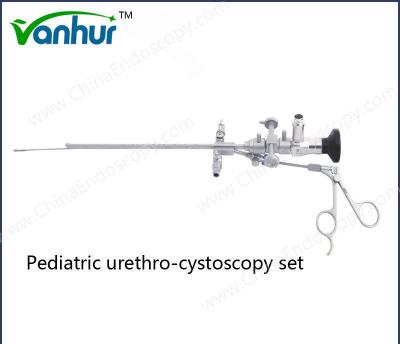
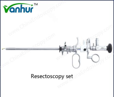
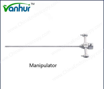
Resectoscopy Electrode Loop Cutting Loop Customization and OEM Options Accepted
1 Introduction:
If you are looking for minimally invasive surgery medical instruments with good quality, competitive price and reliable service. Wanhe medcal is manufaturing these for you. We provide general and professional laparoscopic instruments with CE, FDA approved.
2 Specifications
Adopt 3Cr13, 304, 630 stainless steel material
Tough construction
Corrosion resistant
High durability
Safety application
3 Packing & Shipping:
Package detail: | Poly bag and special shockproof paper box. |
Delivery detail: | By air |
FAQ
What are the methods for evaluating and tracking the surgical results of urological surgical instruments?
The methods for evaluating and tracking the surgical results of urological surgical instruments mainly include the following aspects:
Statistical methods: Using statistical methods to measure and compare the postoperative effects is one of the key factors in evaluating the surgical effect. Through clinical results analysis, experiments and other means, the surgical effect can be objectively evaluated and analyzed.
Physiological index monitoring: including changes in physiological parameters before and after surgery, such as blood pressure, heart rate, respiration, etc. These indicators can reflect the patient's physiological state, help doctors understand the effect of the operation, and provide a basis for further treatment.
Complication evaluation: The Dindo-Demartines-Clavien grading system is one of the most appropriate tools for judging the results of surgical operations. By evaluating the incidence and severity of complications, the effects of different treatment methods can be compared.
Image fusion and electromagnetic tracking technology: The urinary tumor puncture surgical navigation system based on multi-mode image fusion and electromagnetic tracking combines the accuracy of CT/MR positioning, the real-time imaging function of 4D ultrasound volume data and the high precision of electromagnetic tracking. It uses CT/MR-4D ultrasound fusion and electromagnetic tracking to track the puncture target and puncture needle respectively, so as to achieve precise operation of urinary tumor puncture surgery.
Information management: The application of the full-process traceability information system in the management of operating room instruments can conduct full-process traceability management of surgical instruments and improve the efficiency and safety of surgical instruments.
Medical failure mode and effect analysis (HFMEA): The application of medical failure mode and effect analysis (HFMEA) to manage surgical instruments in urological endoscopic surgery can effectively reduce iatrogenic damage, improve surgical treatment effects, and ensure patient safety.
Robot-assisted biopsy technology: Robot-assisted biopsy technology can track and locate surgical instruments during surgery, and use automated intelligent guidance of the puncture process under the guidance of the navigation system through human-computer dialogue, simplify the operation steps, and improve surgical accuracy and efficiency.
In summary, the surgical outcome evaluation and tracking methods of urological surgical instruments cover statistical evaluation, physiological index monitoring, complication evaluation, image fusion and electromagnetic tracking technology, information management, medical failure mode and effect analysis, and robot-assisted biopsy technology, aiming to improve surgical results, reduce complications, ensure patient safety and improve surgical efficiency.
What are the specific applications and effect evaluation of statistical methods in urological surgery?
In urological surgery, the application and effect evaluation of statistical methods are mainly reflected in the following aspects:
Statistical analysis of indicators such as operation time and intraoperative bleeding:
In laparoscopic urological upper urinary tract surgery, the effects of different schemes can be evaluated by analyzing indicators such as operation time, intraoperative bleeding, drainage tube removal time, and postoperative hospitalization time through statistical methods. For example, Liu Sai's study showed that there was no significant difference between the ERAS and CRAS groups in these indicators (P>0.05).
Similar statistical analysis was also conducted for the application of 3D laparoscopic system in urological surgery, including operation time, intraoperative blood loss, postoperative gas discharge time, postoperative extubation time, and postoperative hospital stay days.
Evaluation of patient satisfaction and quality of life:
Patient satisfaction is one of the important indicators reflecting the effect of surgery. By investigating and analyzing patient satisfaction, we can understand the patient's satisfaction with the surgical results and find out the existing problems to improve them.
Analysis of the application of continuous care in the placement of double J tubes in urological surgery showed that all indicators of the observation group were better than those of the control group, with statistical significance (P<0.05), indicating that continuous care can improve patients' self-management ability, quality of life and service.
Evaluation of long-term effects:
The evaluation of the long-term effect of hypospadias surgery needs to focus on the patient's urination function, appearance and function in adulthood, so as to comprehensively evaluate the success rate of the operation and quality of life.
Evaluation of the effect of specific surgical methods:
The effect evaluation of holmium laser enucleation of the prostate (HoLEP) was summarized and the surgical effect was evaluated by retrospectively analyzing the surgical data of 40 consecutive patients.
The evaluation of the postoperative effect of ureteral reconstruction adopts modified upper urinary tract imaging urodynamics to explore its value in guiding subsequent treatment.
The intervention effect of the concept of rapid recovery combined with psychological care:
Statistical methods such as t-test and χ2 test are used to evaluate the impact of the concept of rapid recovery combined with psychological care on patients undergoing urological surgery. The results show that this method can improve patients' self-efficacy, prevent postoperative complications, reduce postoperative pain, promote postoperative recovery of patients, and improve the quality of life.
How to accurately evaluate the recovery after urological surgery through physiological indicator monitoring?
The following methods can be used to accurately evaluate the recovery after urological surgery through physiological indicator monitoring:
Observe surgical indicators and postoperative recovery status: According to the study in the Journal of Clinical Urology, the postoperative recovery of patients can be evaluated by comparing the operation time, intraoperative blood loss, gastrointestinal function recovery time, eating time, bed activity time, and tube removal time of the two groups of patients.
Use the Postoperative Quality Recovery Scale (Pqrs): During the recovery process after general anesthesia and surgery, the Postoperative Quality Recovery Scale (Pqrs) is used to measure the recovery quality and patient satisfaction. This approach takes into account factors such as the type of surgery, care, inflammation, anesthetic drugs and techniques, patient comorbidities, and different views on good recovery.
Application of postoperative rating scales: The application of postoperative rating scales can help doctors understand the patient's recovery progress, predict rehabilitation prognosis, evaluate the effectiveness of rehabilitation treatment, and provide a basis for the development of rehabilitation plans.
Rapid recovery surgical care: The use of rapid recovery care can effectively reduce postoperative complications and improve the quality of life of patients. For example, in a study, the observation group used rapid recovery care and the control group used conventional care. The results showed that the postoperative recovery-related indicators and nursing satisfaction of the observation group were better than those of the control group.
Physiological assessment: Although it is mainly used in coronary PCI treatment, physiological assessment technology can also be used to monitor recovery after other surgeries. Longitudinal vascular analysis of the stent segment can help the surgeon clearly understand the presence of residual blood flow-restricting lesions, thereby optimizing the surgical process.
Pain assessment and care: Pain assessment and care after adult surgery is also an important aspect. The visual analog scale (VAS) is a commonly used subjective assessment method that can effectively assess pain intensity by asking patients to mark the degree of pain on a 10cm straight line.
How accurate and reliable is the Dindo-Demartines-Clavien grading system in the evaluation of complications in urological surgery?
The accuracy and reliability of the Clavien-Dindo-Clavien grading system in the evaluation of complications in urological surgery have been widely recognized and applied. This system can accurately reflect the severity of complications, and its conclusions are consistent with the conventional evaluation criteria in their respective fields, thus having strong advantages in evaluating the safety of surgery, comparing different surgical methods, and analyzing the learning curve of surgical techniques.
Specifically, the Clavien-Dindo grading system has been applied to a variety of urological surgeries, such as transurethral resection of the prostate (TURP), bipolar plasma resection of the prostate (PKRP), and holmium laser enucleation (HoLEP). The system can be used to standardize the differences in complications of these procedures. In addition, the system has also been used to grade early complications after ureteroscopic holmium laser lithotripsy, and the influencing factors of early postoperative complications have been explored.
Nevertheless, some studies have pointed out that the Clavien-Dindo grading system still needs to be further improved and developed to ensure its consistency and accuracy in dynamic clinical settings. However, overall, the system has been proven to be an objective and reproducible outcome assessment tool and has been widely accepted and used in multiple surgical specialties such as urology.
What are the studies on the accuracy and safety of image fusion and electromagnetic tracking technology in urological tumor puncture surgery?
The studies on the accuracy and safety of image fusion and electromagnetic tracking technology in urological tumor puncture surgery mainly focus on the following aspects:
Multimodal image fusion technology, such as MRI-TRUS fusion, mpMRI-TRUS fusion, etc., has been widely used in prostate cancer puncture biopsy. These technologies improve the accuracy of positioning by fusing different types of medical images (such as magnetic resonance imaging and ultrasound images). For example, Carbon Medical VENUS multimodal image fusion ultrasound system has significantly improved the positive detection rate in clinical applications, and has good patient tolerance and high puncture safety.
The electromagnetic positioning system uses the principle of electromagnetic induction to determine the position and positioning information by placing sensors in the patient's body or on surgical instruments and measuring the changes in electromagnetic signals. This system usually has high positioning accuracy and is suitable for complex surgical environments. For example, the urology team of Zibo Hospital of Integrated Traditional Chinese and Western Medicine used magnetic resonance-ultrasound image fusion for transperineal prostate puncture biopsy. Through freehand 3D modeling with a dual-plane ultrasound probe, the registration was completed in 2 minutes with high speed and high precision, shortening the operation time.
Many clinical studies have shown that the puncture surgery navigation system combining multimodal image fusion and electromagnetic tracking technology can improve the accuracy and safety of surgery. For example, the Affiliated Cancer Hospital of Fudan University conducted a retrospective analysis of the data of 50 patients who underwent prostate puncture biopsy after simultaneous mpMRI and 68 Ga-PSMA PET/CT examinations, and explored the effect of mpMRI and 68 Ga-PSMA PET/CT image fusion to guide prostate targeted puncture biopsy.
Although multimodal image fusion and electromagnetic tracking technology have significant advantages in improving the accuracy and safety of urinary tumor puncture surgery, there are also some challenges. For example, although optical navigation has high positioning accuracy, it is easily affected by visual occlusion and is only suitable for open surgery. Therefore, the selection of an appropriate navigation method requires comprehensive consideration based on specific surgical conditions and needs.
What are the latest advances in robot-assisted biopsy technology in improving the precision and efficiency of urological surgery?
Robot-assisted biopsy technology has made significant progress in improving the precision and efficiency of urological surgery. Here are a few key recent advances:
Robot navigation-prostate puncture biopsy: Xinhua Hospital Urology Department successfully carried out "robotic navigation-prostate puncture biopsy". This technology combines the patient's nuclear magnetic resonance (MRI) images with the robot system to achieve more accurate prostate puncture biopsy.
Minimally Invasive® Robot: Minimally Invasive® Robot has demonstrated its application results in high-difficulty surgeries in the field of urology, assisting clinical experts in achieving a number of breakthroughs with important clinical value and milestone significance. In addition, the Tumai® single-arm laparoscopic surgical robot completed the first domestically produced single-arm robotic urological surgery at Shanghai Changzheng Hospital, further improving the accuracy and safety of the surgery.
Magnetic resonance-color Doppler ultrasound fusion prostate puncture biopsy technology: An innovative robot-assisted magnetic resonance-color Doppler ultrasound fusion prostate puncture biopsy technology has been introduced, providing a more accurate and reliable means for the early diagnosis and treatment of prostate cancer. This technology combines multiple imaging methods to improve the accuracy of biopsy.
MRI/ultrasound fusion navigation prostate puncture biopsy surgery: A hospital in Anhui Province successfully carried out robot-assisted MRI/ultrasound fusion navigation prostate puncture biopsy surgery. This is the first time that this advanced technology has been used for prostate puncture biopsy in the region.
For more photos and details please contact me:
Company Name: Tonglu Wanhe Medical Instruments Co., Ltd.
Sales: Emma
Tel:+86 571 6991 5082
Mobile: +86 13685785706